
 |
|
Chest MR images (1 cm thick slices) of the same normal subject. Left:
fast spin-echo proton image. Right: FLASH He3 image of the gas in
the lungs. The NMR frequency is changed from 64 MHz for protons to
49 MHz for He3. |
 |
|
Dynamic images collected using a sliding window radial sequence; the
time interval between displayed images is 0.5s.
|
 |
|
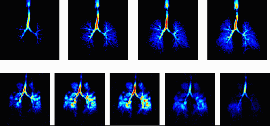
Left to right: gas distribution images, ADC maps and ADC histograms
in 3 subjects (top to bottom).
|
 |
|
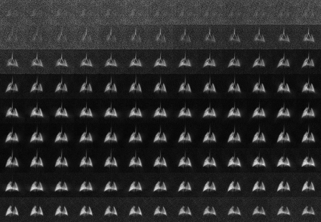
High temporal resolution (100 images/second) of a rat breathing cycle
(20s total acquisition time).
|
 |
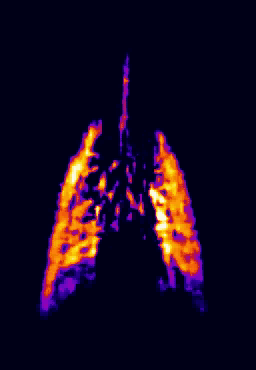 |
| High resolution ventilation image of a rat lung using a dedicated HP
Helium3 respirator. Image: Y. Crémillieux, Lyon |
Dynamic ventilation image series of high temporal resolution in a normal
rat
|
 |
|
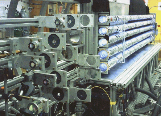
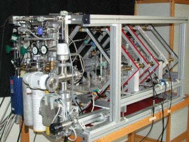
| Optical pumping laboratory setup at Mainz University Images: J. Schmiedeskamp, Mainz |
Photograph of the transport units containing a spherical glass vessel of V = 1 liter. Relaxation times of more than 100 h are routinely achieved in these containers.
|
 |
 |
| Photograph of a compact prototype gas polariser developed in Paris (size:
1.1x0.64x0.64 m3), routinely used for MRI since 2003. Images: PJ Nacher, Paris |
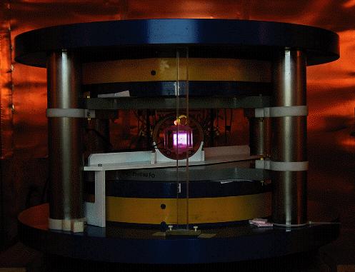
Vertical-axis 3mT whole-body MR system used for in-vivo relaxometry
studies
|
 |
 |
|
Low-field (0.08 T) permanent Magnet developed in the Cracow Group for
small animal MRI.
|
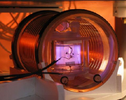
Calibration of the antenna using a low pressure optically pumped cell |
Home
last updated on January
20th, 2006 by P.J. Nacher